A substantially updated version of the hypertextbook is available here. Please migrate to that version. This one will eventually disappear.
LeftBarFeatures of Antimicrobial Tolerance in Biofilms
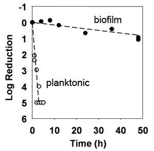
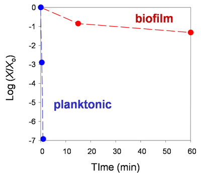
When microorganisms band together in a biofilm they become protected from killing by antimicrobial agents. Three examples of biofilm tolerance, each from a different in vitro laboratory system, are shown in Figures 1, 2, and 3. In each of these graphs, the y-axis is a measure of the surviving number of bacteria on a logarithmic scale. The first example shows the action of an antibiotic, rifampin, on Staphylococcus epidermidis in a colony biofilm (a model that will be discussed further below) (Figure 1).

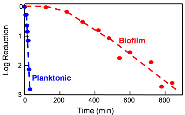
Whereas rifampin kills planktonic bacteria effectively, it has little effect on biofilms even after two days of continuous exposure. Figure 2 shows the action of a common industrial biofilm and medical sterilant, glutaraldehyde, against a biofilm model consisting of Pseudomonas aeruginosa entrapped in gel beads (Grobe et al, 2002). The biofilm is killed more slowly than free-floating bacteria.


These examples and a large literature demonstrate several common features of the tolerance exhibited by microorganisms in biofilms: 1) biofilms formed by diverse bacterial species, and also fungi, manifest tolerance in the biofilm mode of growth; 2) biofilm tolerance has been described for a wide variety of antimicrobial agents with different chemistries and modes of action; 3) antimicrobial tolerance has been reported in a wide variety of biofilms, whether thin or thick, flat or bumpy; 4) the fact that biofilms formed in simple laboratory models also display profound tolerance shows that specific environmental factors (e.g., a cytokine in an animal or humic acids in a surface water) are not essential for the development of reduced susceptibility.
