Harvesting and Dispersing of Cells from Biofilms (Alternative Method)
Instructor Version (go to Student Version)
| Subject Area(s) | microbiology |
| Intended Audience |
high school biology, independent study/science fair, introductory undergraduate microbiology, advanced college level |
| Type | laboratory exercise |
| Revision Date | November 21, 2003 |
CONTENT
This exercise describes methods by which biofilm associated cells may be harvested from slides, coupons1, or other experimental or natural surfaces and separated into single celled suspensions. These dispersed suspensions may be used for purposes of enumeration or for other experimental procedures.PREREQUISITES
Students should be able to define a biofilm, describe the differences between biofilm (surface-attached) and planktonic (free-floating) bacteria, and be able to describe why bacteria usually grow on surfaces. Students should also be familiar with standard methods for serial dilution and plating of microbial cells on standard media. Students must understand the basic premises of bacterial plate counting. It is normally understood that each colony on a plate represents the clonal descendents of a single bacterium. If biofilms are not adequately disaggregated, this assumption may fail. There are a number of methods of disaggregation and some are better than others.INSTRUCTIONAL OBJECTIVE
Given readily available materials, detailed instructions and diagrams, the student will be able to harvest surface attached cells from a biofilm (on a glass slide for example), transfer them to a tube of sterile diluent and disperse them so that they may be used in other laboratory exercises. Students will come to understand that there are various techniques for disaggregating cell masses and that no one technique is best in all circumstances.INSTRUCTIONAL PROCEDURES
- Photographic or diagrammatic images of biofilms will be shown to the students. These should illustrate the complex community in which biofilm cells typically grow. See biofilm images at the ASM MicrobeLibrary at http://www.microbelibrary.org/
- Students will be provided with a complete set of materials required for the harvesting and dispersal of biofilm associated cells along with detailed instructions and diagrams for carrying out this procedure.
- The students will be given an opportunity to review the materials and instructions and to ask questions concerning the procedure.
- Students will be provided with a slide, or other object on which a biofilm has been established and they will carry out the harvesting process.
- The harvested cells will be transferred to a tube containing sterile diluent.
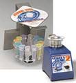
- These tubes are then either turbo mixed or sonicated (2-3 min) to disperse the cells.
- This procedure can then be followed by traditional dilution and plating techniques or by the dilution and drop plating techniques described in another exercise in this collection.
MATERIALS AND EQUIPMENT
Quantity |
Description |
| As Necessary | Wooden applicator sticks, 6 inches long and sterilized by autoclaving (see alternative methods of autoclaving in the Instructors Manual) upright in screw capped tubes [These sticks are used for scraping biofilm from the surface of a slide or coupon (one stick for each student). |
| As Necessary | Sterile culture tubes with caps (18 x 150 mm) |
| As Necessary | Phosphate buffered saline (PBS) |
| As Necessary | Mechanical pipetting devices and sterile pipettes or automatic pipetters, pipettes and pipette tips |
| 1 | Branson or equivalent sonic cleaning water bath |
| 1 | Vortex mixer |
| As Necessary | microscope slides, coupons or other object with biofilm attached3 [An ideal coupon is made by Erie Scientific Company Portsmouth, NH, 800-258- 0834 or www.eriesci.com. These are diagnostic slides with preprinted areas of known dimensions. Using these as coupons, the student can know precisely the area from which cells are being harvested.] |
Directions for making Phosphate Buffered Solution:
To 900 ml of distilled water add 8.0 g sodium chloride, NaCl; 0.2 g potassium chloride, KCl; 0.2 g potassium phosphate, monobasic, KH2PO4; 0.1 g magnesium chloride, hexahydrate, MgCl2.6H2O; and 1.15 g sodium phosphate, dibasic, Na2HPO4.Dissolve completely and add 0.10 g calcium chloride, CaCl2, dissolved in a little water. Adjust to pH 7.4 with either HCl or NaOH as appropriate. Adjust total volume to 1 liter by adding distilled water. Sterilize by autoclaving2
ASSESSMENT / EVALUATION
Assessment may be made by the instructor through a microscopic examination of a simple stained slide (1% aqueous crystal violet). A comparison of a scraped region with an unscraped region of the slide will permit the teacher to evaluate the effectiveness of the student's scraping technique. The teacher could also evaluate the student's scraping technique through the results of associated exercises, e.g. plate count or drop plate count.Teacherís Note:
There is an alternative method of dispersing the cells in the alternative method described here which uses the TURBO MIX™ attachment for a Vortex-Genie mixer. This method is described in the teacherís manual.

FOLLOW-UP ACTIVITIES
This combination of activities can lead to a large number of other exercises, including plate counting, drop plate counting, and any other exercise in which a dispersed population of cells of biofilm origin is required (antimicrobic resistance testing, or the harvesting of cells from contact lens cases, for example).REFERENCES
Development of a Standardized Antibiofilm Test
Zelver, N., M. Hamilton, D. Goeres, D. Walker, and J. Heersink, in Microbial Growth in Biofilms: Part B (R.J. Doyle, Ed.): Methods in Enzymology, Volume 337, pp. 363-376 (2001). See page 366 for scraping and plating techniques.
"Biofilm samples from the rotating disk reactor are obtained by aseptically removing the test coupon from the rotor and removing the biofilm with a sterile wooden applicator stick. The stick is stirred vigorously into a test tube containing 9 ml of sterile buffered water. The entire coupon surface is scraped approximately three times for 1-2 min. The coupon is rinsed with 1 ml of sterile buffered water into the original 9 ml, bringing the final volume of the tube to 10 ml. Prior to enumeration, the cells are disaggregated by homogenization at a speed of 20,500 rpm for 30 sec to eliminate biofilm clumps." pp. 366
A reprint of this paper can be obtained by emailing the Center for Biofilm Engineering, publications@erc.montana.edu. Request paper 01-021
1A coupon is an experimental surface on which biofilms may be grown. The biofilm may then be examined directly by microscopy or sampled for quantification or to determine its properties.
2Standard methods for the examination of water and wastewater : including bottom sediments and sludges 18th ed. ©1992 American Public Health Association, New York.
3Example exercises of how to grow biofilms on objects can be found at http://www.personal.psu.edu/faculty/j/e/jel5/biofilms/. See Buried Slide Technique, Microbial Fishing. This referred site is not maintained by the Biofilm Institute and he Biofilm Institute is not responsible for the site contents.
Educational Program Curricula and Teaching Resources
Supported in part by the Waksman Foundation for Microbiology
Developed in collaboration with Dr. John Lennox, Penn State University-Altoona
© 1999-2008 Center for Biofilm Engineering, http://www.biofilm.montana.edu
