Detachment and Dispersal
Paula Watnick and Roberto Kolter have described biofilms as “microbial cities” with a complex mixture of ethnic neighborhoods, transportation and communication networks, mechanisms which protect the inhabitants from harm and a constant urban renewal program in which removal of some neighborhoods is balanced by the growth of new ones. Being part of a microbial city is important to bacteria for many reasons, not the least of which is that without the attachment to a substratum, the cells would be washed away.
On the other hand, there are times when the ability to leave the city and “get out of town” may be the most favorable alternative. If, for example, nutrient supplies fail or competition becomes too onerous, or your section of the city is suffering the microbial equivalent of urban decay, there may be an advantage to doing what the early pioneers did when the cities became “too crowded” in setting off for new vistas (Figure 1).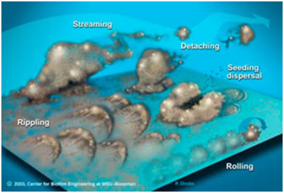
Of course as Watnick and Kolter point out, when you are cemented in place and surrounded by a sticky matrix, the “getting out” part may not be the easiest thing to do. Biofilm specialists have used the terms detachment and dispersal to describe the stages in biofilm development when masses of biofilm material or individual cells may separate from the biofilm and travel to other areas either with the fluid flow and in some cases aided by their own flagellar motion.
Detachment, then is any mechanism which causes individual cells or cell masses to separate from the substratum to which the biofilm is attached. This may include passive forces such as abrasion due to the impact of particles suspended in the fluid flowing past or erosion as fluid flow plucks individual cells from the surface of the biofilm, fluid shear which exceeds the viscoelastic capability of the biofilm or it might represent the actions of the human with a can of detergent and a scrub brush (Figure 1).
Finally there is increasing evidence that biofilms may have the capability to initiate detachment on their own. Enzymes such as alginate lyase coded by the Pseudomonas gene alg L can hydrolyze the matrix encasing the biofilm and cause substantial pieces to detach and many labs have observed what may be called seeding dispersal, a phenomenon which begins as the interior of a biofilm micro-colonies liquefies and ends as the outer layer of the colony is breached permitting the “escape” of the now phenotypically planktonic cells and leaving behind a hollow, empty colony. This process can be particular dramatic in the case of motile species like Pseudomonas aeruginosa because prior to the final lysis of the colony wall, the cells about to be released are seen to be highly motile due to the recent formation of flagellae.

So then, dispersal is facilitated by any mechanism in which “propagules” (fragments or single cells) are caused to leave the existing biofilm with the potential of initiating a new biofilm elsewhere. In the community sense, these are true pioneers (See K vs. r selection).
So, detachment is usually associated with dispersal and dispersal is typically a concomitant event with detachment but there are exceptions.
There are instances in which fragments are removed from biofilms and devoured by predators thus preventing dispersal (B) and there are also dispersal events such as rolling, rippling and swarming motion associated with type IV pili (see Video 2) in which dispersal is not associated with detachment.
In rolling, a roughly spherical mass of cells can be seen to roll down stream in the direction of the bulk fluid flow. The ball is tethered to the substratum and as the ball rolls, new viscoelastic contacts are made while those upstream, stretch and break (Movie). In rippling, the mass of biofilm appears to move in waves propelled at as much as 1 mm/hour by the force of the flowing water. If this doesn’t seem like much, consider that is about 1000 times the length of a typical gram-negative cell each hour.
An understanding of detachment mechanisms has great significance in medicine and industry. The ability to prevent growth of biofilms or alternatively to initiate detachment would save literally billions of dollars in terms of clogging of water pipes, heating and cooling systems, the hulls of ships, fuel delivery systems and in numerous other applications. The ability to do the same in a clinical setting translates not only into lowered health cost, but also in lives saved. The body’s specific and non-specific resistance mechanisms are quite proficient in dealing with planktonic cells but very poor at dealing with biofilm infections. The ability to cause the detachment of a biofilm on a prosthetic hip joint, or on a synthetic heart valve would enable the body’s own defenses to deal with the acute infection caused by the planktonic cells where the biofilm infection might go either undetected by the immune or phagocytic systems of the body or worse serve as a nidus for a vigorous inflammatory response that can cause more damage than the initial infection itself.
In the winter of 1993 and the spring of 1994, hundreds of people became ill with symptoms of acute bacterial pneumonia and before the epidemic resolved itself, one hundred people died. Detailed examination of the medical records of large numbers of these patients revealed two startling facts. All of the patients were asthmatics and all were using the same generic brand of albuterol rescue inhaler.
Albuterol sulfate is a fast acting bronchodialator which when inhaled deeply during an asthma attack, causes the smooth muscle tissue of the airway to relax resulting in improved airflow to the lungs. The rescue inhaler does not cure asthma, and its effects are relatively short term but quite rapid.
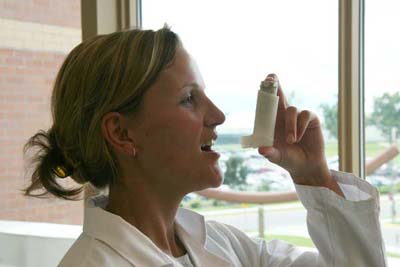
Figure 3. Patient using Albuterol inhaler
Further investigation focused on the manufacturer’s albuterol storage tank. Volumes of albuterol sulfate were prepared and stored in a tank from which the pressurized inhalers were filled prior to distribution. The manufacturer followed industry protocol for cleaning and disinfecting the tank between batches of the drug. But in this case those procedures failed.
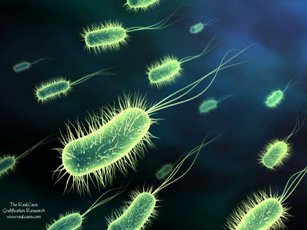
The reason for this failure was the entry into the tank of a very common, usually benign, bacterium called Pseudomonas aeruginosa. This organism is a common inhabitant of soil, water and the surface of plants. It is routinely cultured from the skin of humans and under most conditions is quite harmless. But P. aeruginosa is a bacterial Jekyll and Hyde. The organism possesses two characteristics that make it a threat, particularly to “compromised” individuals such as those with asthma. First, P. aeruginosa is a biofilm former and is capable of growing on animate and inanimate surfaces such as the interior of a storage tank. In this biofilm mode the organisms is nearly impervious to the effects of disinfectants and cleaning agents, accounting for the failure of these agents to remove or kill the organism. Secondly, complementing the mild mannered Jekyll character in the story there is a “malevolent” Hyde. Pseudomonas aeruginosa is called an opportunistic pathogen, that is, given the proper conditions it can cause very serious, even lethal disease.
Biofilms, growing in an environment such as the innocent looking storage tank frequently slough off sizeable masses of material containing bacterial cells and the slimy matrix that normally surrounds them. These fragments were packaged into the inhalers right along with the albuterol. The patients, their airways already compromised by the asthma attempted to inhale the medicine as deeply into their bronchioles as possible, and in this case delivered albuterol and Pseudomonas far into their lungs.
Once there the bacterium forms another biofilm, this time, in the bronchioles and alveoli of the lungs. The organism attaches to the tissue surface, and begins to secrete a sticky, slimy polysaccharides, which surround and protect the bacterium from host antibodies and prowling white blood cells which would normally attack and destroy individual bacteria.
Here, however, the life-saving medicine became the vehicle for a deadly pathogen. William Costerton, a biofilm expert called to testify in some of the many court cases that resulted as a result of this epidemic commented that, “If you are unfortunate enough to aspirate a biofilm [of Pseudomonas] the bacteria have a 100% chance of surviving in your lungs. Good medical treatment and intensive antibiotic therapy saved many of the victims of this tragic accident. One hundred patients were not so fortunate.
Potera, Carol, 1996. Biofilms Invade Microbiology. Science 273: 1795-1797.

